Monday, November 7, 2011

Washington People: Rochelle Smith

DBBS Staff Member Rochelle Smith is featured in this week's RECORD:

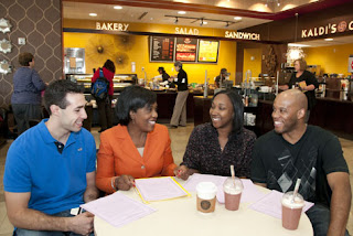
Rochelle Smith (second from left) talks with students (from left) David Cotter, Paige Cooper and Donell Carey, all graduate students in the Division of Biology and Biomedical Sciences, about volunteer opportunities.


Rochelle Smith has a knack for recruiting underrepresented graduate students in the sciences to Washington University.


In the past five years, the number of minority students entering graduate programs each year in the biomedical sciences has almost tripled, as have the number of students entering the Medical Scientist Training Program (MSTP) and the Division of Biology and Biomedical Sciences’ (DBBS) summer research programs for undergraduate students. She travels the country to recruit students and finds ways to bring them to the university so they can see it firsthand.

Smith is director of diversity, summer programs and community outreach for DBBS. She is known for her magnetic personality that puts students, faculty and staff at ease.

“Rochelle represents the best of WUSTL,” says Brian Sullivan, executive director of the university’s MSTP. “She balances her commitment to excellence and effectiveness with a concern for the individual, which results in changes not only to structures but also to hearts and minds.”

READ MORE


Wednesday, October 5, 2011

Natural compound helps reverse diabetes in mice

Researchers (from left) Shin-ichiro Imai, MD, PhD, Jun Yoshino, MD, PhD, and Kathryn Mills showed that a natural compound, NMN, helps to treat symptoms of diabetes in mice. Photo by: Julia Evangelou Strait

Researchers at Washington University School of Medicine in St. Louis have restored normal blood sugar metabolism in diabetic mice using a compound the body makes naturally. The finding suggests that it may one day be possible for people to take the compound much like a daily vitamin as a way to treat or even prevent type 2 diabetes.

This naturally occurring compound is called nicotinamide mononucleotide, or NMN, and it plays a vital role in how cells use energy.

"After giving NMN, glucose tolerance goes completely back to normal in female diabetic mice,” says Shin-ichiro Imai, MD, PhD, associate professor of developmental biology. “In males, we see a milder effect compared to females, but we still see an effect. These are really remarkable results. NMN improves diabetic symptoms, at least in mice.”

The research appears online Oct. 4 in Cell Metabolism.

READ MORE

DBBS Faculty Member wins prestigious Presidential Early Career Award

The White House announced Sept. 27 that Lan Yang, PhD, assistant professor of electrical and systems engineering in the School of Engineering & Applied Science at Washington University in St. Louis, has been named a recipient of the Presidential Early Career Awards for Scientists and Engineers (PECASE).

The early career award is the highest honor bestowed by the United States government on science and engineering professionals in the early stages of their independent research careers. This year, there are 94 recipients.

“It is inspiring to see the innovative work being done by these scientists and engineers as they ramp up their careers — careers that I know will not only be personally rewarding but also invaluable to the nation,” President Barack Obama said in the award announcement. “That so many of them are also devoting time to mentoring and other forms of community service speaks volumes about their potential for leadership, not only as scientists but as model citizens.”

“I am pleased that the President has honored Lan with this special award for her world-class record of achievement,” says Ralph S. Quatrano, PhD, dean of the School of Engineering & Applied Science.

“She is a model for other assistant professors, and I have great confidence in her potential for continued future success. As only the fourth assistant professor at Washington University to receive this prestigious recognition since its inception 15 years ago, this award brings great visibility to Lan and her innovative work and to our school and university,” Quatrano says.


READ MORE

Monday, September 12, 2011

Restoration as science: case of the collared lizard

In a time when a five-year grant is considered a long-term grant, Alan R. Templeton, PhD, a professor of biology in Arts & Sciences at Washington University in St. Louis, has managed to follow some of the species he studies for 10, 20 or even 30 years.


Early in his career he studied parthenogenesis, or virgin birth, in fruit fly populations at a dump and in cactus patches in Hawaii.

“Drosophilia have fast generation times,” he says, “but I studied them for 12 years. And because I followed them for 12 years, I saw patterns I wouldn’t otherwise have seen. In fact, had I not stuck with it so long, I often would have made incorrect conclusions.”

But the fruitfly study is a sprint compared to his lizard work, described in the cover story of the September 2011 issue of Ecology. The Ecology article covers more than 20 years of a 30-year followup monitoring the reintroduction of collared lizards on Ozark glades in 1984. (For the story in pictures, see the slideshow to the right.)

During this time, 1,662 lizards living on 139 glades on three mountains were captured or recaptured 4,545 times. The acknowledgements section of the paper thanks more than 20 people for their help in capturing lizards.

The major revelation of the work was that burning entire mountains and valleys, called landscape-level burning, undid ecological damage that was slowed but not stopped by smaller prescribed burns.

In fact, it allowed the lizards to undertake their own expanded restoration effort without the assistance of worried biologists.

Moreover, burning benefited many species besides the lizards, including a rare fen orchid and fen dragonfly, that were flying under the radar and would probably never have commanded labor intensive restoration efforts on their own.

In short, fire turned restoration from a time-consuming labor-intensive process to one that ran pretty much on its own.

READ MORE

Wednesday, June 29, 2011

Sleep switch found in fruit flies

Rather than count sheep, drink warm milk or listen to soothing music, many insomniacs probably wish for a switch they could flick to put themselves to sleep.


Scientists at Washington University School of Medicine in St. Louis have discovered such a switch in the brains of fruit flies. In a study appearing June 24 in Science, the researchers show that a group of approximately 20 cells in the brains of fruit flies controls when and how long the flies sleep. Slumber induced through this sleep switch was essential to the creation of long-term memory, directly proving a connection between memory and sleep that scientists have long suspected.

“This is exciting because this induced sleep state so far appears to be very similar to spontaneous sleep,” says Paul Shaw, PhD, associate professor of neurobiology. “That means we can manipulate these cells to explore a whole new realm of questions about the purposes of sleep. Such studies might one day lead us to more natural ways of inducing sleep in humans.”


READ MORE

Wednesday, June 8, 2011

Needlemans receive Eliot Society ‘Search’ Award

photo by Joe Angeles
Philip (left at podium) and Sima Needleman accept the Eliot Society's highest honor at its annual celebration on May 11, as Chancellor Mark S. Wrighton looks on. The Needlemans received the “Search” Award, which is given each year to a person or couple to recognize extraordinary dedication and service to the university. The event was held at the Ritz-Carlton Hotel in Clayton.


It is one of the most anticipated rites of spring on campus: friends of Washington University in St. Louis gather to mark another year of generosity from the group of alumni, parents and colleagues who collectively provide the largest amount of unrestricted support to the university through membership in the William Greenleaf Eliot Society.


At this year’s Eliot Society event May 11 at the Ritz-Carlton Hotel, members enjoyed fine dining and camaraderie, heard an address by former Secretary of State James Baker, III and watched as Sima and Philip Needleman received the Society’s “Search” Award for their extraordinary dedication to Washington University.

“This year’s Search awardees, Philip and Sima Needleman, have an enduring association with Washington University, and have served the institution in many important ways,” Chancellor Mark S. Wrighton says. “It was a pleasure to acknowledge their significant and lasting contributions.”

Eliot Society President John Beuerlein introduced Baker, JD, who provided timely insights regarding foreign policy and current events in the Middle East.

READ MORE

Tuesday, May 24, 2011

DBBS Hooding and Recognition Ceremony

On Friday, May 20th awarded 13 PhD students and 17 MD/PhD students their degrees. Here are a few pictures taken on that day. We are so proud of our graduates and wish them the best in all of their future endeavours!


Monday, May 9, 2011

Washington People: Kathryn G. Miller

Kathryn G. Miller, PhD (center), professor and chair of the Department of Biology in Arts & Sciences, examines petri dishes with Julie Morrison (right), senior technician, and Mamiko Isaji, PhD, postdoctoral researcher, both in biology. Miller “cares deeply for the people of Washington University and is especially great with students and the young faculty,” says Erik D. Herzog, PhD, associate professor of biology.

By Nancy Fowler


Her nose habitually buried in a Nancy Drew mystery, little Kathy Miller spent much of her girlhood trying to crack the case.

Today, Kathryn G. Miller, PhD, professor and chair of the Department of Biology in Arts & Sciences, still is playing detective.

With Sherlock Holmes-like intensity, Miller studies cells the way a special agent scrutinizes a crime scene. The adjectives “curious,” “detailed” and “practical” sum up Miller’s modus operandi as she focuses on how certain proteins allow particular cells to perform precise functions.

“That’s the way I look at things,” Miller says. “I’ve got to figure out how they work.”

Love on a cellular level


A Chicago-area middle school teacher sparked the passion that eventually would lead to Miller’s own teaching career. With fond memories, she recalls the contagious enthusiasm of her young seventh-grade science instructor — fortuitously named Mr. Bliss.

“I remember looking at pictures of cells and being incredibly interested in what they were and what they were made up of,” Miller says. “I was kind of a nerd.”

Miller’s love of life sciences took her to Lawrence University in Appleton, Wis., for an undergraduate degree in chemistry and shortly thereafter to Johns Hopkins University School of Medicine, where she earned a doctorate.

It was during postdoctoral work at the University of California, San Francisco, that Miller began studying multicellular organisms. In 1989, she came to WUSTL to teach and continue her research; in 2008, she became chair of the biology department.
Even while overseeing 30 biology faculty members, Miller maintains a keen focus on research.

Her work zeroes in on how cells specialize to perform specific functions in a multicellular organism. Of particular interest is how the cells’ structural proteins contribute to three features and activities: assuming different shapes, connecting with various components and having functional properties critical to their roles.

An important byproduct of her research

READ MORE

Wednesday, March 23, 2011

Scientists grow personalized collections of intestinal microbes

Washington University scientists have grown personalized collections of intestinal microbes in the laboratory. The research sets the stage for identifying new probiotics and evaluating whether microbe transplants can restore the natural balance of intestinal bacteria in “sick” microbial communities.


Each of us carries a unique collection of trillions of friendly microbes in our intestines that helps break down food our bodies otherwise couldn’t digest.


This relationship between humans and their microbes is generally a healthy one, but changes to the mix of microbes in the digestive tract are suspected to play a role in obesity, malnutrition, Crohn’s disease and other ailments.

Now, scientists at Washington University School of Medicine in St. Louis have shown they can grow and manipulate personalized collections of human intestinal microbes in the laboratory and pluck out particular microbes of interest.

The research sets the stage for identifying new probiotics and evaluating in preclinical trials whether microbe transplants can restore the natural balance of intestinal bacteria in “sick” microbial communities.

The research, by Jeffrey I. Gordon, MD, the Dr. Robert J. Glaser Distinguished University Professor and director of the Center for Genome Sciences & Systems Biology, and his team is reported online March 21 in the early online edition of the Proceedings of the National Academy of Sciences.


READ MORE

Wednesday, March 9, 2011

Brain Inspector

 height=
Photo by: Michael C. Purdy

Simeon Schlaggar examines a human brain at NeuroDay March 5 at the Saint Louis Science Center as his father, Bradley Schlaggar, MD, PhD, the A. Ernest and Jane G. Stein Associate Professor of Neurology, looks on. Simeon is also the son of Christina Lessov-Schlaggar, PhD, research assistant professor of psychiatry. The event, jointly sponsored by Washington University and the Saint Louis Science Center, was an opportunity for the public to learn about brain science and was staffed in part by Washington University graduate students. Other activities included using a brain-computer interface to play a video game, operating a model of nerve cell communication that used jelly beans and light switches, making a model nerve cell from pipe cleaners, and participating in exercises that highlighted the way the brain analyzes sensory data. NeuroDay was part of International Brain Awareness Week, created by the DANA Alliance for Brain Initiatives and the Society for Neuroscience

Monday, February 14, 2011

Peter Burgers: Biochemist studies cell processes fundamental to life

By Julia Evangelou Strait


Photo by: Robert Boston
Peter Burgers, PhD (center), shows Joseph Stodola (left) and Justin Sparks, both graduate research assistants in biochemistry and molecular biophysics, some samples in his lab. “Peter is known around the world for his expertise in the genetics and biochemistry of DNA replication and cellular responses to damaged DNA,” says Thomas E. Ellenberger, DVM, PhD, the Raymond H. Wittcoff Professor and head of the Department of Biochemistry and Molecular Biophysics. “He is well-known for reconstituting impossibly difficult protein complexes and showing the field how things work at the molecular level.”

Growing up in Wassenaar, Netherlands, a small town near the North Sea, Peter Burgers, PhD, remembers a carefree childhood. The middle of seven children, Burgers and his siblings spent much time outside, including summers at the beach.

But a carefree childhood does not mean one without direction.

“My father was a teacher, and he valued education,” Burgers says. “He wanted all of us to go to university, which was unusual in those days.”

That parental influence clearly was felt as the family now includes a biochemist, an engineer, two medical doctors, two nurses and a geologist. Today, Burgers is the Marvin A. Brennecke Professor of Biological Chemistry at Washington University School of Medicine in St. Louis. He is an expert in DNA replication and repair — fundamental cellular processes shared across organisms, from yeast to humans.

“Peter is known around the world for his expertise in the genetics and biochemistry of DNA replication and cellular responses to damaged DNA,” says Thomas E. Ellenberger, DVM, PhD, the Raymond H. Wittcoff Professor and head of the Department of Biochemistry and Molecular Biophysics. “He is well-known for reconstituting impossibly difficult protein complexes and showing the field how things work at the molecular level.”

Indeed, Burgers recently was honored for his scientific contributions with an honorary doctorate in medicine from UmeĂĄ University in Sweden. During the ceremony, Bengt JĂĄrvholm, MD, PhD, dean of the UmeĂĄ School of Medicine, praised Burgers’ work, his support for international collaborations, and his mentoring of faculty at UmeĂĄ University. The celebration included Burgers’ general lecture to the UmeĂĄ community. Afterward, festivities continued with a university-wide reception and banquet.


READ MORE

Friday, February 4, 2011

Research scientists urge universities to improve undergraduate science teaching

On Jan. 12, a bleak commentary in Nature online suggested a funding bubble in the sciences is about to collapse with dire results for science students. “When the economic storm struck in 2008, the ride came to an abrupt end,” wrote Colin Macilwain, a contributing correspondent at Nature.


“As Western governments attempt to maintain investment in science as a route to innovation and industrial development, they are undermining support for students and the quality of their education,” Macilwain said.

“Students are not customers of a university; they are its very soul,” Macilwain wrote. “The idea that research will prosper while teaching and learning decay is a dangerous fallacy.”

Two days later, in the Jan. 14, 2011, issue of Science, Sarah C.R. Elgin, PhD, the Viktor Hamburger Distinguished Professor in Arts & Sciences at Washington University in St. Louis, and 12 colleagues took issue with Macilwain’s fundamental premise.

Elgin — a professor of biology and of education in Arts & Sciences and of biochemistry and molecular biophysics and of genetics in the School of Medicine — and her colleagues don’t believe teaching takes place at the expense of research or research at the expense of teaching.

She and her Science co-authors write that “excellence in scientific research and teaching need not be mutually exclusive but are instead intertwined and can interact synergistically to increase the effectiveness of both.”

The authors are biomedical research scientists who receive support from the Howard Hughes Medical Institute to create “new programs that more effectively engage students in learning science.” So, they admit that their situations are more favorable than most.

Elgin comments as well that Washington University has the resources to fund programs out of the reach of state colleges, even in this time of financial hardship.

READ MORE

Thursday, February 3, 2011

New nanoparticles make blood clots visible

Wiley-VCH Verlag GmbH & Co. KGaA
A blood vessel (top) with ruptured atherosclerotic plaque, shown in yellow, is developing a blood clot. The nanoparticles, shown in blue and black, are targeted to a protein in the blood clot called fibrin, shown in light blue. A traditional CT image (bottom left) shows no difference between the blood clot and the calcium in the plaque, making it unclear whether this image shows a clot that should be treated. A spectral CT image (bottom right) “sees” the bismuth nanoparticles targeted to fibrin in green, differentiating it from calcium, still shown in white, in the plaque.


For almost two decades, cardiologists have searched for ways to see dangerous blood clots before they cause heart attacks.


Now, researchers at Washington University School of Medicine in St. Louis report that they have designed nanoparticles that find clots and make them visible to a new kind of X-ray technology.

According to Gregory Lanza, MD, PhD, a Washington University cardiologist at Barnes-Jewish Hospital, these nanoparticles will take the guesswork out of deciding whether a person coming to the hospital with chest pain is actually having a heart attack.

“Every year, millions of people come to the emergency room with chest pain. For some of them, we know it’s not their heart. But for most, we’re not sure,” says Lanza, a professor of medicine. When there is any doubt, the patient must be admitted to the hospital and undergo tests to rule out or confirm a heart attack.

“Those tests cost money and they take time,” Lanza says.

Rather than an overnight stay to make sure the patient is stable, this new technology could reveal the location of a blood clot in a matter of hours.

READ MORE

Monday, January 31, 2011

New method takes snapshots of proteins as they fold

Scientists have invented a way to ‘watch’ proteins fold — in less than thousandths of a second — into the elaborate twisted shapes that determine their function.


By Diana Lutz


An unfolded protein is a string made up of amino acids. When the protein folds it forms structures like the alpha helix (the corkscrew) and the beta sheet (the flat ribbon) in the image to the right. These secondary structures then double back on themselves to form the final structure.

People have only 20,000 to 30,000 genes (the number is hotly contested), but they use those genes to make more than 2 million proteins. It’s the protein molecules that do most of the work in the human cell. After all, the word protein comes from the Greek prota, meaning “of primary importance.”

Proteins are created as chains of amino acids, and these chains of usually fold spontaneously into what is called their “native form” in milliseconds or a few seconds.

A protein’s function depends sensitively on its shape. For example, enzymes and the molecules they alter are often described as fitting together like a lock and key. By the same token, misfolded proteins are behind some of the most dreaded neurodegenerative diseases, such as Alzheimer’s, Parkinson’s and mad cow disease.

Scientists can’t match the speed with which proteins fold. Predicting how chains of amino acids will fold from scratch requires either powerful supercomputers or cloud sourcing (harnessing the pattern recognition power of thousands of people by means of games such as Folding@home).

Either way, prediction is time-consuming and often inaccurate, so much so that the protein-structure bottleneck is slowing the exploitation of DNA sequence data in medicine and biotechnology.

A clever way of watching proteins fold and unfold may finally provide the kind of detail needed to improve protein structure predictions.


READ MORE

Thursday, January 27, 2011

Biologists’ favorite worm gets viruses

Marie-Anne Felix, the Monod Institute

Scientists have discovered that C. elegans, a microscopic worm biologists have used in the lab to identify important biological phenomena, suffers from natural viral infections. This may mean that C. elegans can help scientists learn more about how hosts and viruses interact.

A workhorse of modern biology is sick, and scientists couldn’t be happier.

Researchers at Washington University School of Medicine in St. Louis, the Jacques Monod Institute in France and Cambridge University have found that the nematode C. elegans, a millimeter-long worm used extensively for decades to study many aspects of biology, gets naturally occurring viral infections.

The discovery means C. elegans is likely to help scientists study the way viruses and their hosts interact.

“We can easily disable any of C. elegans’ genes, confront the worm with a virus and watch to see if this makes the infection worse, better or has no effect,” says David Wang, PhD. “If it changes the worm’s response to infection, we will look to see if similar genes are present in humans and other mammals.”

READ MORE

Wednesday, January 26, 2011

Cancer information tool for journalists wins Health 2.0 developer challenge

Health 2.0 and the National Cancer Institute (NCI) recently named Ozioma, an online cancer information tool from the Health Communication Research Laboratory (HCRL) at Washington University in St. Louis, one of two winners of a national contest.


The Ozioma News Service was chosen a winner of the Enabling Community Use of Data for Cancer Prevention and Control Challenge, a part of the 2010 Health 2.0 Developer Challenge.

The Ozioma tool helps reporters and media relations professionals create localized cancer stories for specific populations in specific communities.

View a brief video about Ozioma.

“For those who write about and report on health, providing a central location to access community-level health data can increase the likelihood that they’ll include these data in their stories,” says Charlene A. Caburnay, PhD, research assistant professor at the Brown School at Washington University.

READ MORE

Tuesday, January 25, 2011

Three DBBS faculty receive Children’s Discovery Institute grants

Three Washington University School of Medicine researchers have received grants from the Children’s Discovery Institute to advance their work into childhood diseases.


The Children’s Discovery Institute is a partnership between Washington University School of Medicine and St. Louis Children’s Hospital to accelerate better treatments, cures and preventions for childhood diseases.

Todd Druley, MD, PhD, received a CDI Faculty Scholar Award for his research into the genetic basis of a fast-growing cancer of the white blood cells called high-risk acute lymphoblastic leukemia (ALL) that primarily affects teenagers .


Druley
Through his work in the Center for Genome Sciences and Systems Biology, Druley developed a method in 2009 for surveying many genes in a pool of DNA from more than 1,000 people using next-generation sequencing. This work was published in Nature Methods. Now, he is applying that method to children with high-risk ALL looking for genetic mutations that may be behind the cancer.

“It is unlikely that there will be one or two mutations in DNA that cause children to get ALL,” he says. “We predict that there will be a lot of different mutations in a lot of different genes that have little consequence individually, but can have a synergistic effect that results in a serious problem.”

By targeting genes already identified from genome-wide arrays done on children with high-risk ALL, Druley will study several hundred genes in about 450 DNA samples from children enrolled in nationwide high-risk ALL trials.

High-risk leukemia affects about one in 100,000 children, but unlike standard-risk ALL, is curable only about 65 percent to 75 percent of the time. Unfortunately, physicians haven’t made much progress in treating the disease with chemotherapy, so many patients require bone marrow transplants, which can be difficult physically and emotionally for these teens.

“We hope to understand how high-risk ALL is coming about in the first place so we can design more effective treatments,” says Druley, an assistant professor of pediatrics and of genetics at the School of Medicine who also treats patients at St. Louis Children’s Hospital.

Druley began the work as a postdoctoral researcher in the Washington University lab of Robi Mitra, PhD, assistant professor of genetics, in the Center for Genome Sciences and will continue the work with the CDI grant through at least 2013.

Kelle Moley, MD, has received a grant from the Children’s Discovery Institute to facilitate research among Washington University researchers on pregnancy, maternal-fetal interaction and pediatric diseases.

The project, the Women and Infants Health Specimen Consortium (WIHSC), will collect tissue samples such as cord blood and amniotic fluid from mothers and their infants and link them with a clinical database. Participants will be recruited from the Washington University Reproductive Endocrinology and Infertility Clinic and the Center for Advanced Medicine. This project is unique, Moley says, because individual mother-infant pairs will be followed before, during and after pregnancy.

“Washington University has many researchers who study women and infants’ health, disease, fertility, pregnancy and the neonatal period,” says Moley, the James P. Crane Professor of Obstetrics and Gynecology. “The pooling of resources could lead to increased access to a collection of specimens from pregnant and nonpregnant women, which would undoubtedly help facilitate research.”

Samples collected through the WIHSC will be available to School of Medicine investigators to study areas such as fetal and developmental origins of childhood diseases; the interaction and communication between the fetus and mother in utero; and to possibly identify biomarkers that predict poor pregnancy and poor infant outcomes, such as miscarriage and pre-term births.

“This consortium has the potential to become a vital source of tissue and patient data, from both mother and child, needed to accelerate new pathways to discovery in childhood disease,” Moley says.

Ann Gronowski, PhD, associate professor of pathology and immunology, and Marwan Shinawi, MD, assistant professor of pediatrics, are co-principal investigators of the WIHSC.


Sheila Stewart, PhD, received a grant to expand a library of molecular tools that can selectively turn off every gene in the mouse genome.

Called an shRNA library, it is a resource that allows researchers to “knock down” or deplete a gene of interest. By turning off a gene and observing the consequences, scientists can gain insight into that gene’s role in the biological process. This library will provide the Washington University research community with the tools to study human disease in a mouse model at a much lower cost.

Deleting or inserting a gene and observing the consequences is not a new research technique. While this technology has changed the way researchers ask important questions, standard methods can be cumbersome, time-consuming and expensive. An shRNA library, however, takes advantage of RNA interference (RNAi), a more recently discovered mechanism for turning off the expression of genes. Normally, a gene codes a strand of messenger RNA, which then codes for a protein. Traditionally, scientists would remove the gene, eliminating its messenger RNA and the resulting protein. With RNA interference, instead of going through the difficult process of removing the gene, scientists use shRNA strands that interfere with the messenger RNA and prevent it from creating the protein.

“Because of the way we created this system, we can introduce our RNAi construct to any kind of cell,” Stewart says. “This approach uses the RNAi machinery that is naturally in cells to take out the gene we tell it to. Once there is none of that gene’s protein left, we can look at the function of the protein in the context of the disease we’re studying. Did removing the protein make the disease better or worse or change anything?”

Elaine Mardis, PhD, co-director of The Genome Center and director of technology development, and David Piwnica-Worms, MD, PhD, professor of developmental biology and of radiology, are collaborating with Stewart on the CDI grant.

Researchers at The Genome Center, led by Mardis, also associate professor of genetics and of molecular microbiology, are making DNA that is usable for researchers studying disease, and Stewart's group is turning it into an infectious virus that can be used to screen for genes that impact a wide variety of human diseases.

It’s very expensive to make usable DNA and introduce it into a cell, Stewart says, but by turning it into an infectious virus, researchers can get it into almost all cells. These libraries represent an opportunity for WUSTL researchers to study any type of cell, especially those most relevant to the disease a researcher is interested in, Stewart says.

Stewart’s lab is also training other researchers on how to use the library. Earlier, Stewart received a CDI grant to expand the human genome library.

Monday, January 24, 2011

Deep genomics: Scientists probe the epigenome

The Washington University in St. Louis team participating in the modENCODE project, a massive ongoing effort to map all the elements in living cells that affect whether genes are expressed or silenced, discuss a fruit fly strain with an enzyme that affects gene expression. Team members are (from left): Sarah C.R. Elgin, PhD, the Viktor Hamburger Distinguished Professor in Arts & Sciences who led the team; Sarah Gadel, lab technician; Nicole C. Riddle, PhD, research assistant professor of biology in Arts & Sciences; and Tingting Gu, PhD, a postdoctoral research associate in biology. Missing is Sarah Marchetti, a technician. A major paper that includes their part of the modENCODE project has just been published in Nature.


In 2003, the year a complete draft of the human genome was released, the U.S. National Human Genome Research Institute launched the ENCODE project (ENCyclopedia of DNA Elements), to develop an encyclopedia of the epigenome, that is, of all of the many factors that can change the expression of the genes without changing the genes.


Four years later, the National Institutes of Health funded modENCODE (the Model Organism ENCylopedia of DNA Elements) to work out the epigenomes of two model organisms: the fruit fly Drosophila melanogaster, lurker among rotten bananas, and the round worm, Caenorhabditis elegans, slitherer between crumbs of soil.

This was big science with a vengeance. The work had been divided among 21 teams, themselves each made up of multiple labs, all toiling away at universities and companies across the country. And that’s not counting the research groups tasked with coming up with new ways of handling the floods of data pouring out of the labs.

What was going on?

Sarah C.R. Elgin, PhD, the Viktor Hamburger Distinguished Professor in Arts & Sciences, who led the Washington University in St. Louis lab that is part of one of the modENCODE teams, offers an explanation.

READ MORE

Friday, January 21, 2011

Interviews Have Begun!

Today marked the start of the 2011 Interview Season. After receiving well over 1000 applications, DBBS will be interviewing hundreds of students for positions in our 12 programs.
For a list of interview weekends, you can go HERE
For information on the Division, click HERE
Dr. John Russell speaking to applicants.

Wednesday, January 19, 2011

Kidney gene implicated in increased heart failure risk

Scientists have identified the first DNA sequence variant common in the population that is not only associated with an increased risk of heart failure, but appears to play a role in causing it.

The variant, a change in a single letter of the DNA sequence, impairs channels that control kidney function.

“It’s not a heart gene,” says Gerald W. Dorn II, MD, the Philip and Sima K. Needleman Professor of Medicine at Washington University School of Medicine in St. Louis and a lead investigator on the study. “It’s a kidney gene. This protein is not even expressed in the heart. Nobody has previously considered that kidney-specific gene defects might predispose you to heart failure.”

Heart failure is diagnosed when the heart can no longer provide sufficient blood to the body. It can have a number of causes, including high blood pressure, cancer therapy, viral infections of the heart or heart attack.

“It’s a syndrome,” Dorn says. “You’ve had sufficient damage to your heart that it doesn’t work very well. You collect fluid in your lungs, you swell up, and you have trouble breathing.”

The unexpected results highlight the advantage of performing genome-wide studies to find DNA sequence variants associated with disease.

READ MORE